

A century after the EEG was discovered, it remains a crucial tool for understanding the brain
By Giridhar Kalamangalam, University of Florida
Jena, Germany, 1924: Working in near-isolation and with painstaking tediousness, the psychiatrist Hans Berger observes rhythmic electrical activity from the scalp of human subjects. He is convinced the activity arises from within the brain and coins the term “electroencephalogram.”
It is 10 years before the scientific community accepts Berger’s work, birthing the field of electroencephalography, or EEG for short.
Today, the electroencephalogram – also abbreviated as EEG – is widely known as a medical test measuring brain electrical activity that’s used on patients who have, or are suspected to have, neurological disorders. The EEG provides a window into the living brain, with a continuous electrical readout of what is happening inside our heads. The procedure may be short, often just a 30-minute recording. But for patients monitored for diagnosis or treatment of brain disease, it can be continued for much longer – days or even weeks.
As a neurologist specializing in epilepsy, I use EEG on a daily basis. Our team at the University of Florida interprets thousands of EEGs a year in neurological patients. I also run a research lab where our goal is to understand the basic structure of the EEG in health and disease.
A history of unexpected twists
The story of EEG is colorful and littered with fables. Berger’s interest in brain electricity was not to combat disease, though that was his day job as a physician, but to find a biological basis for his belief in telepathy. He wondered whether the brain waves of EEG could convey thoughts through space, allowing people to read each other’s minds. He was unsuccessful in his mission, but the field he founded nonetheless took off.
By the mid-1930s, researchers had observed the striking differences between the awake and asleep EEG. The EEGs of patients with brain disease turned up a variety of unprecedented observations.
And then came a defining moment for modern medicine. In December 1934, a group of Boston physicians observed the rhythmic EEG spike-wave appearance of seizures in patients with “petit mal” epilepsy. Petit mal is an anachronistic term for a type of epilepsy where a patient’s flow of thought, speech or action momentarily freezes during seizures. For the first time, the symptoms and behavior of patients during seizures were correlated to a brain signal occurring in lockstep.
EEG quickly evolved from a scientific curiosity to a mainstream clinical tool. The first clinical EEG laboratory was set up at Massachusetts General Hospital in 1937. The practice grew in the ensuing decades into the specialized services that institutions like ours have offered since the 1970s.

The EEG explained
So what, exactly, is the EEG?
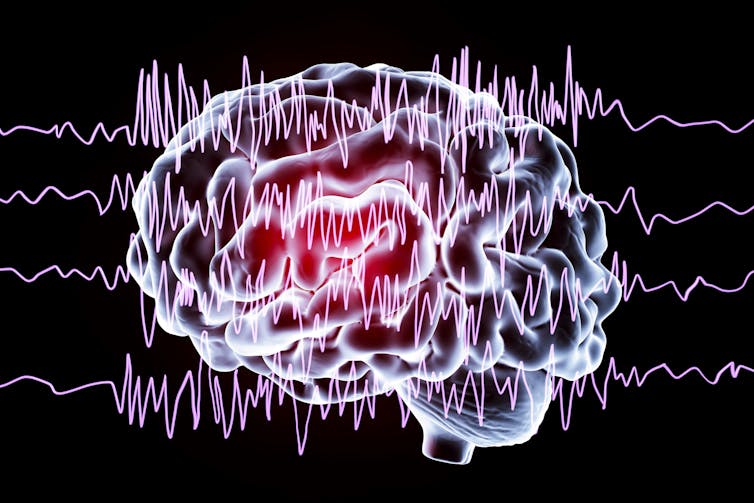
Imagine taking two small metallic discs connected by a conducting wire. Place one disc on the scalp and connect the other to a neutral reference, such as the ear. Watch a tiny alternating current flow in the wire, proportional to the electrical activity sensed by the conducting contact. This activity is the EEG, the electrical milieu that bathes brain tissue.
In turn, the EEG arises from the excitable nature of nerve cells, or neurons. When neurons fire, action potentials – brief, high-voltage spikes traveling outward from their cell bodies – set up local electrical activity in other neurons, causing current to flow within and outside them.
These local current flows may cause the targeted neurons to fire in turn and set up yet more current flows. Thus, the system sustains itself. The average overall activity is a mix of many different frequencies, with the five main ones called delta, theta, alpha, beta and gamma waves.
If the EEG were just random up-and-down drift – “the bloodless dance of action potentials,” commented a skeptical early 20th century neurologist – it would be much less interesting. The remarkable fact is that EEG tends to spontaneously organize into patterns in time and space.
The spike-wave pattern of petit mal, referenced earlier, is a classic example, but scores of others are now known. Clinical EEG practice is just recognizing characteristic EEG patterns and correlating them to specific disease states.
Fluctuating neurons
Beyond the clinic, an unsettling scientific question emerges. Simply put, how do electrical patterns arise in the brain? How do the billions of neurons and their trillions of local current flows fluctuate in just the right ways to create globally ordered structure?
Our research group has been interested in the fundamental question of pattern formation in EEG. It turns out that activity in the brain is naturally repetitive – that is, oscillatory. This is due to the way neurons are connected and the fact that they interact by excitation and inhibition to produce push-pull effects.
Considering local oscillations as fundamental building blocks, we showed that the EEG over the entire brain could be built up from such elementary blocks. More interestingly, the differing frequencies could be made to coalesce, or synchronize, into a common rhythm. We recognized that synchronization of this type underlies some seizurelike patterns observed in patients.
EEG, AI and the mind
Pattern formation in nature is deeply fascinating. How does a leopard get its spots? How does the audience at a concert spontaneously settle into a rhythmic applause? Many such questions trace their origin to a classic paper on biological pattern published in 1952. Its author was Alan Turing, better known as the father of computer science and early advocate of artificial intelligence, or AI.
The hardware underlying the majority of today’s AI systems are neural networks. Neural networks were introduced in 1943 by Warren McCulloch, a physician and electroencephalographer. Like Berger, McCulloch’s interest in EEG extended beyond brain disease. He wondered where in the brain’s neurons and EEG lay the capacity to think. He conceived the idea of grouping artificial neuronlike computing units into networks, analogous to how real neurons in the brain interconnected.
Together with Walter Pitts, he proved that such neural networks could function as a general-purpose computer. The seminal McCulloch-Pitts ideas were refined over the ensuing decades and reside in the “deep learning” neural networks of today’s AI.
Deep learning AI has infiltrated all areas of biomedicine, including neurology. For example, AI systems can successfully interpret brain scans. AI methods have also been used to analyze EEG.
Can AI systems be trained to deduce thoughts from the EEG? Can AI approach Berger’s quest for telepathy? Incredibly, recent deep-learning AI research has shown that some aspects of mental activity may be decoded from EEG.
In 2024, EEG turns 100. What windows will it open into the brain and mind in the future? Doubtless, clinical applications will grow. Surely, brain pattern generation will be better understood. Perhaps EEG will glimpse the content of the mind. And for neurologists like me surveying the AI revolution, there’s the quiet pride that EEG was really at the start of it all.![]()
Giridhar Kalamangalam, Professor of Neurology, University of Florida
This article is republished from The Conversation under a Creative Commons license. Read the original article.